Tag Archives: genome stability

rDNA theory of aging

The ribosomes are the most abundant protein complexes in the cell. They are synthesised from ribosomal DNA (rDNA) genes within nucleoli. In most eukaryotic cells, rDNA exists in tandem arrays, which are repeating units of similar sequences. Oftentimes the rDNA provide sites for recombination, and the copy number seems to fluctuate. In this regard, the rDNA is hypothesised to be involved in genome stability and cellular senescence.

There  was a paper written by Takehiko Kobayashi last year, 2008, in the journal BioEssays, wherein he proposed the rDNA theory of aging, stating that the rDNA may be the region most sensitive to DNA damage. He highlighted two key molecular entities that would link rDNA to aging: FOB1 and SIR2. These two are well known aging genes.

“… in yeast Saccharomyces cerevisiae … lifespan expansion in a fob1 mutant is associated with increased rDNA stability (no copy number variation), while the lifespan reduction in a sir2 mutant is associated with decreased rDNA stability (high copy number variation) …”

“rDNA is more unstable and induces checkpoint control faster than any other part of genome, and that the nucleolus may be more sensitive to protein damage because it is protein-dense …”

He stated further that rDNA may serve as ‘‘sensor’’ for DNA damage, and also as ‘‘shock absorber’’ that protects the genome from damage. I think these are broad statements as of this time.

He and his colleagues have observed that fob1 mutants show better growth than wild type, indicating checkpoint induction is reduced and lifespan is extended. More specifically, he mentioned that rDNA copy number gradually decreases in a fob1 mutant by a FOB1-independent form of homologous recombination. This implies termination of growth, presumbaly because of a shortage of rRNA and ribosomes. In this manner, lifespan extends due growth delay or termination. And yet, the number of abnormal cells is likely to increase.

In contrast, he described that sir2 mutants show increased rDNA instability. He stated that this leads to greater checkpoint control and shorter lifespan. I shall look further into this as it seems unclear to me.

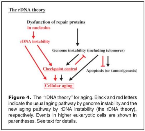
Below is one of the images he presented in the paper:

rdnatheory

A new role of the rDNA and nucleolus in the nucleus – rDNA instability maintains genome integrity
Takehiko Kobayashi
National Institute of Genetics and The Graduate University for Advanced Studies, SOKENDAI, 1111 Yata, Mishima 411-8540 Japan
email: Takehiko Kobayashi (takobaya@lab.nig.ac.jp)
BioEssays (2008)
Volume 30 Issue 3, Pages 267 – 272

Leave a comment

Filed under ageing

BLAP75/Rmi1

Liudmila Chelysheva and colleagues studied BLAP75/Rmi1 in relation to BLM/Sgs1 and TopoIIIα/Top3. Their paper appeared on the December 2008 issue of PLoS Genetics journal. Below is the summary:

“Recombination is a process by which cells can repair DNA damage. Such repair can either be crossovers (CO), in which DNA molecules are submitted to major exchanges, or non-crossover (NCO) events. Eukaryotic cells have developed several mechanisms to maintain genome stability during vegetative development by limiting the occurrence of CO events in favour of NCO. BLAP75/Rmi1, BLM/Sgs1, and TopoIIIα/Top3 act together in a complex (BTB/RTR) known to be a crucial component of regulation mechanisms against CO formation. However, CO/NCO regulation is thought to be very different during meiosis since homologous chromosomes (paternal and maternal) overcome at least one CO/pair. In this study, we investigate the role of the BTB/RTR complex during meiotic recombination through the analysis of a function of one of its members: the A. thaliana homologue of BLAP75/Rmi1. We show for the first time that BLAP75/Rmi1 is also a key protein of the meiotic homologous recombination machinery. In Arabidopsis, we found that this protein is dispensable for homologous chromosome recognition and synapsis, but necessary for the repair of meiotic double-strand breaks. Furthermore, in the absence of BLAP75, bivalent formation can happen even in the absence of CO.”

Chelysheva L, Vezon D, Belcram K, Gendrot G, Grelon M. 2008. The Arabidopsis BLAP75/Rmi1 Homologue Plays Crucial Roles in Meiotic Double-Strand Break Repair. PLoS Genet 4(12): e1000309. doi:10.1371/journal.pgen.1000309

Note: A. thaliana blap75 mutants are sterile. They examined the reproductive development of these mutants and found that blap75 sterility is due to abortion of male and female gametophytes. In particular, blap75 mutants show defects in male sporogenesis.

Leave a comment

Filed under DNA damage, DNA repair, genetics, genome stability

ATR and H2AX

Last month (December 2008)  in the Journal of Biological Chemistry, Rebecca Chanoux and colleagues reported the results of their studies on ATR and H2AX. They wrote:

“If ATR prevents the collapse of stalled replication forks into DSBs, and H2AX facilitates HR-mediated restart, the combined deficiency in ATR and H2AX would be expected to dramatically enhance the accumulation of DSBs upon replication fork stalling. Herein, we utilize both partial and complete elimination of ATR and H2AX to demonstrate that these genes work cooperatively in non-redundant pathways to suppress DSBs during S phase.”

They hypothesized that H2AX participates in a Rad51-mediated suppression of DSBs generated in  the absence of ATR.

They found that increased Rad51 focal accumulation in ATR-deficient cells is dependent on H2AX, and deficiencies in  both ATR and H2AX lead to synergistic increases in chromatid breaks and translocations.

They discussed further that the ATM and DNA-PK phosphorylation site on H2AX (Ser139) is required for genome stabilization in ATR’s absence; therefore, phosphorylation of H2AX by ATM and DNA-PKcs plays a pivotal role in suppressing DSBs during DNA synthesis in instances of ATR pathway failure.

Their results imply that fork stabilization (ATR-dependent) and H2AX/ATM/DNA-PKcs-dependent pathways cooperate to suppress DSBs when replication stalls.

Leave a comment

Filed under ATR, biochemistry, DNA, DNA damage, DNA repair, double strand break, genetics, genome stability, H2A.X, homologous recombination, recombination

telomere loss produces genomic instability

Simon Titen and Kent Golic studied telomere loss by breakage of an induced dicentric chromosome in the fruitfly Drosophila melanogaster [Genetics 2008 Dec;180(4):1821-32; Epub 2008 Oct 9]. They found that one outcome of this is cell death through Chk2 and Chk1 controlled p53-dependent apoptotic pathways. Yet they also observed escape from apoptosis and repeated division in some cells that have lost a telomere. This evasion of apoptosis is accompanied by abnormalites such as chromosome fusions, anaphase bridges, aneuploidy, and polyploidy. They also found evidence of bridge–breakage–fusion cycles and chromosome segments without centromeres. They further noted that cells manifesting these signs of genomic instability were much more frequent when the apoptotic mechanisms were crippled. In the end, they concluded that the loss of a single telomere is sufficient to generate genomic instability involving multiple chromosomes and aneuploidy.

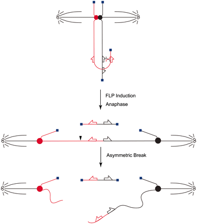
They used the FLP recombinase system to examine the cellular responses to loss of a single telomere. This generates a chromosome end lacking a telomere. Synthesis of FLP recombinase is induced by heat shock, which causes recombination between inverted FRTs on sister chromatids to generate dicentric and acentric chromosomes. They believe their strategy closely mimics the situation faced by a mammalian cell in which a single telomere becomes critically short and dysfunctional due to incomplete replication in the absence of telomerase.

011

Figure 1. Dicentric/acentric chromosome production and segregation. FLP-induced recombination between oppositely oriented FRTs on sister chromatids produces a dicentric chromosome and an acentric chromosome. At anaphase the dicentric chromosome is stretched between the poles and usually breaks. Centromeres are indicated as filled circles, telomeres as filled squares, FRTs as arrows.

Leave a comment

Filed under biochemistry, cell biology, chromosomal defect, DNA damage, drosophila, FLP recombinase, genetics, genome, genome stability, molecular biology, recombination

perinuclear tethering promotes rDNA repeat stability

Karim Mekhail and colleagues studied a network in the the budding yeast Saccharomyces cerevisiae that stabilizes ribosomal DNA repeats thru interactions between rDNA-associated silencing proteins and proteins of the inner nuclear membrane (INM).

They reported that deletion of either the INM or silencing proteins:

—reduces perinuclear rDNA positioning

—disrupts the nucleolus–nucleoplasm boundary

—induces the formation of recombination foci

—destabilizes the rDNA repeats

“In addition, artificial targeting of rDNA repeats to the INM suppresses the instability observed in cells lacking an rDNA-associated silencing protein that is typically required for peripheral tethering of the repeats.

Moreover, in contrast to Sir2 and its associated nucleolar factors, the INM proteins are not required for rDNA silencing, indicating that Sir2-dependent silencing is not sufficient to inhibit recombination within the rDNA locus.

These findings demonstrate a role for INM proteins in the perinuclear localization of chromosomes and show that tethering to the nuclear periphery is required for the stability of rDNA repeats.”

[for more details]

Their results suggest that Sir2-dependent silencing alone cannot inhibit recombination within the repetitive rDNA locus and that INM-mediated perinuclear chromosome tethering ensures repeat stability.

They expect that the proteins studied are members of perinuclear networks that control recombination at multiple loci to maintain genome stability.

Leave a comment

Filed under ageing